
Have you started noticing glare while driving at night, blurred vision, halos around lights, or difficulty seeing clearly in bright conditions? These symptoms can sometimes be associated with a cortical cataract.
A cortical cataract is a type of cataract that develops in the outer portion, or cortex, of the eye’s natural lens. It can progress gradually, and the effect on vision depends partly on whether the cloudy areas extend toward the central visual axis.
This complete guide explains what cortical cataract means, its causes and risk factors, common symptoms, stages and grading, treatment options, and when surgery may be considered.
What Is a Cortical Cataract?
A cortical cataract develops in the outer layer, or cortex, of the eye’s natural lens. Unlike nuclear cataracts, which affect the lens centre, cortical cataracts typically begin at the periphery and form characteristic spoke- or wedge-shaped opacities that may extend inward.
These changes can scatter light as it passes through the lens, often causing glare, halos, and reduced contrast sensitivity, particularly in bright conditions or while driving at night.
Cortical cataracts are one of the main types of age-related cataracts and can occur alongside other cataract types.
How Common Are Cortical Cataracts?
Cortical cataracts account for approximately 15-25% of all age-related cataracts — making them the second most common type after nuclear cataracts. They are significantly more prevalent in:
- Women than men — particularly post-menopausal women
- Patients with Type 2 diabetes — where altered glucose metabolism accelerates cortical fibre disruption
- Individuals with significant lifetime UV-B exposure — outdoor workers, those at high altitude or low latitude
- Adults over 60 — prevalence increases steeply with each decade after 60
5 Major Causes of Cortical Cataract
Cause 1 — Age-Related Cortical Fibre Degeneration
The most common cause — progressive breakdown of the cortical lens fibres that maintain ion transport and water balance within the lens. As these fibres lose their structural integrity with age, water accumulates in clefts between fibres, disrupting protein arrangement and creating the characteristic cortical opacities.
- Affects the majority of adults over 60 to some degree
- Progression rate varies widely — some cortical cataracts remain stable for years
- Associated with reduced antioxidant enzyme activity in ageing lens tissue
Cause 2 — Ultraviolet B (UV-B) Radiation Exposure
UV-B radiation from sunlight is one of the strongest environmental risk factors specifically for cortical cataract — more strongly associated with cortical than nuclear cataract formation.
- Cumulative lifetime UV-B exposure drives photooxidative damage to cortical lens cells
- Outdoor workers, agricultural workers, and those without UV-protective eyewear are at greatest risk
- UV400 sunglasses have been consistently shown to reduce cortical cataract risk in epidemiological studies
Cause 3 — Diabetes Mellitus
Diabetes is one of the major metabolic drivers of cortical cataract — chronically elevated blood glucose disrupts the osmotic balance within cortical lens fibres through sorbitol accumulation and altered sodium-potassium pump function.
- Diabetic patients develop cortical cataracts earlier and with faster progression
- Poorly controlled blood sugar accelerates cortical opacity formation significantly
- Well-controlled HbA1c measurably delays diabetic cortical cataract progression
Cause 4 — Nutritional Deficiencies
Deficiencies in Vitamin C, Vitamin E, lutein, and zeaxanthin — the lens’s primary antioxidant defences concentrated in the cortical layer — reduce protection against oxidative damage that drives cortical opacity.
- Vitamin C is particularly concentrated in the aqueous humour surrounding the lens cortex
- Low dietary antioxidant intake is consistently associated with higher cortical cataract prevalence
- Supplementation may slow progression when dietary intake is inadequate
Cause 5 — Steroid Medication and Other Drugs
Long-term corticosteroid use — systemic or topical — is associated with posterior subcapsular cataracts primarily but also contributes to cortical opacity in some patients through altered lens metabolism.
- Certain diuretics (thiazides) are independently associated with cortical cataract risk
- Phenothiazine medications produce characteristic anterior cortical deposits
- Patients on long-term medication regimens should have regular eye examinations
Symptoms of Cortical Cataract
Cortical cataract symptoms reflect the optical effect of the spoke-like opacities scattering light as it enters the eye — particularly from peripheral light sources:
Primary symptoms:
- Glare and halos — the most characteristic symptom; oncoming headlights, sunlight, and overhead lighting are particularly disabling
- Reduced contrast sensitivity — difficulty distinguishing objects from backgrounds of similar tone
- Blurred vision — initially in peripheral vision, worsening as opacities extend centrally
- Monocular diplopia — double vision or ghost images in the affected eye when tested alone
- Difficulty with night driving — the combination of glare and reduced contrast makes nighttime driving especially problematic
Important distinction from nuclear cataract: Cortical cataracts produce disproportionate glare and contrast reduction relative to measured visual acuity — a patient may have reasonably good Snellen acuity but be significantly disabled by glare in real-world lighting conditions.
Risk Factors for Cortical Cataract
Beyond the five primary causes, these factors independently increase cortical cataract risk:
- Females— women have consistently higher cortical cataract prevalence in large population studies
- Myopia — short-sightedness is associated with increased cortical cataract risk
- Low socioeconomic status — associated with greater lifetime UV exposure and poorer nutritional status
- Hypertension — independently associated with cortical opacity in several cohort studies
- Alcohol consumption — chronic heavy drinking depletes lens antioxidant reserves
- Dark iris colour — paradoxically associated with slightly higher cortical cataract risk in some studies
Types of Cortical Cataracts
Cortical cataracts are classified by their morphological pattern and location within the cortex:
Anterior cortical cataract: Opacities in the front layer of the cortex adjacent to the anterior capsule. Often the earliest cortical changes detected on slit lamp examination — may remain stable for extended periods.
Posterior cortical cataract: Opacities in the rear cortical layer adjacent to the posterior capsule. More visually significant than anterior cortical changes because of proximity to the optical centre.
Equatorial cortical cataract: The most characteristic pattern — spoke-like or cuneiform opacities beginning at the lens equator (periphery) and extending radially toward the centre. This is the classic “cortical cataract” appearance.
Lamellar (zonular) cortical cataract: A discrete zone of cortical opacity surrounding a clear nucleus — often congenital or early-onset, associated with metabolic disturbances during a specific developmental period.
Stages of Cortical Cataract Progression
| Stage | Description | Visual Impact |
| Incipient | Small peripheral spoke-like opacities at the lens equator; does not reach visual axis | Minimal — glare may be noticed in specific lighting |
| Immature | Opacities extend further centrally; some clear cortex remains | Moderate — glare, reduced contrast, mild blurring |
| Mature | Complete cortical opacity; no clear cortex remaining | Significant visual impairment |
| Hypermature | Cortical liquefaction; potential complications | Severe — urgent surgical consideration |
Most cortical cataracts remain in the incipient to immature stage for many years before significant functional impact develops — making regular monitoring important for timely surgical planning.
Cortical Cataract Treatment Options
Non-Surgical Management (Early Stage)
In the incipient and early immature stages, when visual function remains adequate:
- Updated spectacle prescription — optimising remaining visual acuity through glasses
- Anti-glare coatings — particularly beneficial for cortical cataract patients whose primary complaint is glare
- UV400 sunglasses — crucial for slowing further UV-driven cortical progression
- Nutritional support — Vitamin C, lutein, and zeaxanthin supplementation where dietary intake is inadequate
- Blood sugar and metabolic optimisation — for diabetic patients, strict HbA1c control measurably reduces progression rate
Important: No medication, eye drop, or supplement reverses established cortical cataract opacity. These measures support the remaining clear lens tissue and slow further development — they do not cure existing opacities.
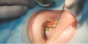
Surgical Treatment — Phacoemulsification
When cortical cataract reaches the stage of functional visual impairment, phacoemulsification is the definitive treatment:
Cortical cataracts and phacoemulsification: The soft, water-containing nature of cortical cataract material is actually advantageous for surgery — the lens nucleus is typically softer than in advanced nuclear cataracts, requiring less ultrasound energy and producing less stress on the corneal endothelium.
IOL options for cortical cataract patients:
| IOL Type | Best Suited For |
| Monofocal IOL | Maximum reliability; glasses for distance or near |
| Toric IOL | Patients with significant corneal astigmatism |
| Multifocal / Trifocal IOL | Spectacle independence goals |
| EDOF IOL | Extended range with reduced glare profile |
When Is Surgery Required for Cortical Cataract?
The decision to proceed with phacoemulsification for cortical cataract is based on functional impact — not the appearance of the cataract alone:
Surgery is indicated when:
- Glare significantly impairs driving safety — particularly night driving
- Best corrected visual acuity has fallen below 6/18 in the affected eye
- Contrast sensitivity reduction is measurably limiting daily function
- The patient’s occupational or lifestyle visual demands are not being met with optical correction
- The cortical cataract has progressed to the mature stage
Surgery can be deferred when:
- Visual acuity remains above 6/9, and glare is manageable
- The patient’s daily visual demands are low
- Medical comorbidities require optimisation before elective eye surgery
Risks and Complications of Cortical Cataract Surgery
Phacoemulsification for cortical cataract is exceptionally safe — but informed consent includes:
Cortical-specific surgical considerations:
- Posterior capsule rupture — cortical cataracts with dense cortical material require careful aspiration technique
- Residual cortical material — incomplete cortical cleanup can cause post-operative inflammation (uveitis)
- Capsular phimosis — the anterior capsule can contract excessively post-operatively in some cortical cataract cases
Standard cataract surgery risks (all types):
- Posterior capsule opacification (PCO) — secondary cataract treatable with YAG laser capsulotomy
- Endophthalmitis — very rare intraocular infection
- Cystoid macular oedema — post-operative macular fluid causing temporary blurring
How to Prevent Cortical Cataract
While age-related cortical changes cannot be entirely prevented, these evidence-based strategies meaningfully delay onset and slow progression:
- UV400 sunglasses consistently outdoors — the single most effective environmental prevention measure for cortical cataract specifically
- Strict blood sugar control — particularly important for diabetic patients where HbA1c directly influences cortical progression rate
- Antioxidant-rich diet — leafy greens (lutein, zeaxanthin), citrus (Vitamin C), nuts and seeds (Vitamin E)
- Stop smoking — reduces systemic oxidative burden on the lens from the first month of cessation
- Annual eye examinations from age 50 — or earlier if risk factors are present, so progression is monitored, and surgery is timed optimally
- Minimise unnecessary steroid use — only under ophthalmological supervision with regular slit lamp monitoring
Frequently Asked Questions
1. Is a Cortical Cataract Serious?
A cortical cataract can become significant when it interferes with vision and everyday activities. Its impact varies between people, and an eye examination can determine its severity and whether treatment is needed.
2. What Is the Difference Between a Cortical and Nuclear Cataract?
A cortical cataract affects the outer cortex of the lens, often causing spoke-like opacities and glare. A nuclear cataract affects the central nucleus and may cause progressive blurring, yellowing or hardening of the lens. Both types can occur together.
3. Are Cortical Cataracts Easy to Remove?
Cortical cataracts can normally be treated effectively with cataract surgery when they become visually significant. The complexity of surgery depends on the cataract and the overall health of the eye.
4. What Is the Most Serious Type of Cataract?
There is no single cataract type that is considered the most serious for every patient. The impact depends on the cataract’s location, severity, effect on vision, and the presence of other eye conditions.
5. Do Cataracts Ever Get Better Without Surgery?
An established cataract does not normally clear on its own or with eye drops and supplements. Updated glasses may improve vision temporarily in early stages, but cataract surgery is the definitive treatment when the cataract significantly affects vision.
Conclusion
Cortical cataracts can develop gradually, often causing glare, blurred vision, and difficulty seeing clearly in certain lighting conditions. Timely Evaluation helps determine the cataract’s severity and whether monitoring, updated glasses, or surgery may be appropriate.
At AG Vision, patients can receive a comprehensive cataract evaluation and discuss treatment options based on their individual eye health.
For patients seeking cataract care in Noida, Dr. Arushi Garg can assess the condition and explain suitable treatment options, including cataract surgery when clinically appropriate.